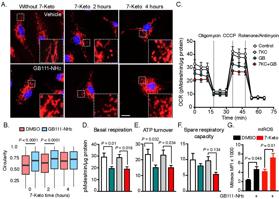
Fig. 5. Cathepsin inhibition causes mitochondrial dysfunction and alters its networked structure. Time resolved microscopy of mitochondrial structure in response to 7-Keto. (A) BMDMs were treated with GB111-NH2 or DMSO for 16 h and then received 7-Keto or vehicle at the indicated time points. BMDMs were stained with mitotracker deep red for 30 min and fixed in cold methanol prior to microscopy examination (scale bar is 10 µm). (B) Mitochondrial circularity is presented in a box and whisker plot. One-way ANOVA with Tukey's post hoc test was used to compute statistical significance. The data represent a summary of three independent experiments. (C-F) Live cell mitochondrial-metabolic performance was measured using a Seahorse metabolic analyzer. BMDMs were cultured in assay plates and treated with GB111-NH2 or DMSO as described in (A). 7-Keto was added two h before the metabolic assay. Real time oxygen consumption was measured by the Seahorse machine and respiratory performance was assessed as described in the Materials and Methods. Basal respiration, ATP turnover and spare respiratory capacity rates were inferred from the curve, normalized to total protein content and calculated as described in the Materials and Methods. Each assay contained 4-6 biological replicates, measured in three technical replicates. The data are summarized over 7 independent experiments. One-way ANOVA with Sidak's post hoc test was used to evaluate statistical significance. (G) Flow cytometry analysis of mitochondrial reactive oxygen species (mtROS) production. BMDMs were treated with GB111-NH2 or DMSO as described in (A) and stimulated with 7-Keto for another 4 h. Mitosox was used to evaluate mitochondrial ROS levels and measured by flow cytometry. One-way ANOVA and Tukey's post hoc test were used to compute statistical significance from three independent experiments. Bar graphs present the mean ± SEM.